2026-09-26 · 21 min · connectomics · drosophila · neuroscience · datasets · open-data · explainer

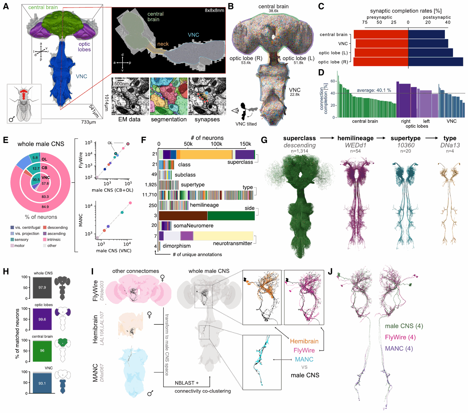
On September 25, Google posted that researchers from Google Research and HHMI Janelia had built "the first complete brain map for a male fruit fly", and that "we mapped every single neural connection in a male fruit fly brain and central nervous system, amounting to more than 166,000 neurons." The paper behind the post is Berg et al., "Sexual dimorphism in the complete Drosophila male central nervous system connectome", Cell 189, 5504–5526, published September 3, 2026 (doi:10.1016/j.cell.2026.08.015). It is open access under CC BY. The preprint went up in October 2025 (doi:10.1101/2025.10.09.680999), and the dataset, MaleCNS, reached v1.0 on June 8, 2026.
I read the Cell paper, the preprint's methods and supplementary figures, Google's post, Janelia's release and the dataset pages, and read the volume's metadata from the public bucket. Every number is labelled reported (from those sources), measured (read off the data by me) or reasoned (my arithmetic).
The short version: the first connectome of a whole male fly central nervous system, brain, optic lobes and nerve cord in one animal. 166,700 neurons, 11,710 cell types, an estimated 44 person-years of proofreading (reported). Read "every single neural connection" carefully. Essentially every traceable neuron was proofread, but 40.1% of detected connections have a proofread neuron at both ends (reported), which is normal for the field.
What a connectome is
A connectome is a wiring diagram at the resolution of single synapses. You image a nervous system with an electron microscope at a few nanometres per voxel. You trace every neuron through the volume. You find every chemical synapse and record which neuron sits on each side. Out comes a directed graph: neurons are nodes, synapse counts are edge weights, and each neuron carries annotations such as cell type, developmental lineage and a predicted neurotransmitter.
It is anatomy. It fixes who can talk to whom, and through how many contacts. It does not measure how strong a synapse is, how fast a neuron integrates or what a neuromodulator does; the paper says functional experiments must confirm what the wiring predicts. That gap is why a model built on a connectome needs a scrambled-wiring control. oruk.ai's speech classifier used 499 neurons from this dataset, ran that control, and the scrambled wiring scored the same.
How this one was built
Eight stages turn one fly into one graph. Machines image, segment and detect synapses. People do most of the rest, and people are the expensive part. The prose after the explorer carries the same numbers.
A gallium ion beam mills 8 nm off a slab face and a scanning electron beam images the fresh surface; repeat. Seven machines ran in parallel for about 13 months.
- voxel
- 8 × 8 × 8 nmreported
- image volume
- 160 teravoxelsreported
- volume / of which tissue
- 0.082 / 0.054 mm³reported
- raw size at one byte per voxel
- about 160 TBreasoned
- orphan linking41%
- neuron review28%
- cleaving14%
- focused merging8%
- cell-body fibre linking5%
- backbone tracing4%
One fly, 66 slabs
The team dissected hundreds of five-day-old males (from a cross of an isogenized Canton S strain with w1118) and kept each central nervous system in one piece: brain, neck connective and ventral nerve cord (VNC, the insect equivalent of a spinal cord). After fixation and staining, 44 candidates went through X-ray CT to look for subtle damage and uneven staining. One sample, Z0720-07m, was imaged (reported).
A whole fly CNS is too large to mill as one FIB-SEM block. So a "hot-knife", a heated, oil-lubricated diamond knife, cut it into 20 µm slabs: 31 transverse slabs through the nerve cord, then 35 sagittal slabs through the brain (reported). Slabs also let seven microscopes work in parallel.
Seven microscopes, 13 months, 160 teravoxels
Focused-ion-beam scanning electron microscopy (FIB-SEM) is a block-face method. A gallium ion beam (15 nA, 30 kV) mills a nominal 8 nm layer off the slab face. A scanning electron beam (3 nA, 1.2 kV landing energy, 3 MHz) images the fresh surface. Repeat until the slab is gone. Because milling depth matches pixel size, voxels are isotropic, 8 × 8 × 8 nm. A fleet of seven enhanced FIB-SEM machines imaged the 66 slabs over 13 months and produced 160 teravoxels covering 0.082 mm³, of which 0.054 mm³ is tissue (all reported). At one byte per voxel, that is about 160 TB of raw image (reasoned; the volume is stored as uint8).
FlyWire's female brain took the other route, serial-section TEM: sections 35–40 nm thick imaged at 4 nm in-plane, so 4 × 4 × 40 nm voxels. It is faster. The FlyWire paper notes that its lower z resolution and more frequent artefacts once made reconstruction harder, which the hemibrain team cited to justify FIB-SEM's cost.
Stitching a neck
Slabs are stitched to their neighbours, and the brain is rotated 18 degrees, head tilted back, to square it with the volume axes (reported). The hard part is the neck. Brain and cord were separated by one cut and sectioned in orthogonal directions, so their surfaces met at about 25 degrees. The fix was one non-rigid transform that fades with distance from the neck. It had to stay local because proofreading had already started in the brain (reported).
I read the aligned volume's metadata from the public bucket:
curl -s https://storage.googleapis.com/flyem-male-cns/em/em-clahe-jpeg/info
# scale 0: resolution [8, 8, 8] nm, size [94088, 78317, 134576], data_type uint8
That bounding box is about 992 teravoxels, roughly 753 × 627 × 1,077 µm (measured size, reasoned product). The imaged 160 teravoxels fill about 16% of it (reasoned); a brain and a cord do not pack into a cuboid.
Segmentation: flood-filling networks
Google's contribution is the automated segmentation, with flood-filling networks (FFNs, Januszewski et al., 2018): a 3D convolutional network that starts at a seed voxel and grows one object's mask, deciding voxel by voxel whether the object continues. In the hemibrain and MANC pipelines this one follows, FFNs first produce a deliberately over-segmented base, and further FFN passes agglomerate it into candidate neurons. (Google's post also mentions PATHFINDER, a newer system; the paper names FFNs for this volume.)
Segmentation ran three times, on the right hemisphere, the left hemisphere and the nerve cord, in the order the volume was assembled (reported). The cord needed its own model to tell muscle from nervous tissue. Merges across each seam were chosen by shared voxel counts, and constraints forbade merges between nuclei and fibres in the same nerve bundle, so automated merges could not corrupt segments already proofread.
Synapses and transmitters
Fly synapses are polyadic: one presynaptic T-bar faces several postsynaptic densities. One network detects T-bars, a second finds their partners, both fine-tuned on this sample. Validation used 114 densely labelled cubes of 300 × 300 × 300 voxels across 81 regions, with 2,303 T-bars and 16,870 postsynaptic partners (reported). The lamina's cubes exposed poor recall on oddly shaped T-bars, so the lamina got its own detector.
The detector found 46 million presynapses connected to 312 million postsynapses, with precision 0.82 and recall 0.81 for a synapse counted as a unit, at a confidence threshold of 0.5 (reported). That is about 6.8 postsynapses per T-bar (reasoned); the hemibrain reported 6.7.
Neurotransmitters are predicted, not measured. A ResNet50 looks at a small EM cube centred on each T-bar (640 nm, in the paper's description) and scores 7 transmitters; predictions are pooled per neuron and per cell type. A consensus column overrides them with experimental ground truth where it exists and marks octopamine and serotonin unclear for lack of validation data (reported).
44 person-years of proofreading
Automated segmentation still splits neurons and merges neighbours, and fixing that is most of the cost. 29 expert proofreaders worked for 3 years, an estimated 44 person-years from edit logs and task records (reported). The protocols:
- Cell-body fibre linking: attach orphaned cell bodies to their neurites.
- Cleaving: split false merges in every significant segment.
- Backbone tracing: coarse-trace synapse-rich segments.
- Focused merging: the tool proposes a merge, a human says yes or no.
- Orphan linking: trace a loose fragment to its backbone.
- Neuron review: inspect whole neurons, often beside their left-right partner.
Excluding scripted edits, that came to 4,117,544 merges, 147,212 cleaves and 6,046 supervoxel splits (reported). Every fragment with 100 or more synaptic connections was traced; in the central brain and right optic lobe the threshold was 50.
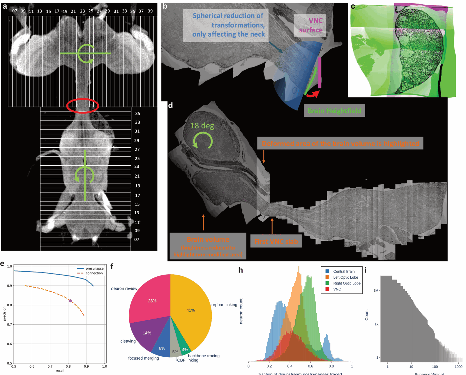
The pie is the useful part. Orphan linking took 41% of the labour and neuron review 28%; backbone tracing, which produces the recognisable shapes, took 4% (reported). Orphan linking alone is about 18 person-years (reasoned). Small fragments are where the time goes.
So the team trained a model, AutoProof, on the yes/no answers from focused merging and ran it on unexamined orphans with 10 or more synapses. Above a conservative threshold, targeting an estimated 3% error rate, merges were accepted without a human: about 200,000 of them, adding about 2.4 million postsynapses, 309,000 T-bars and 1.3% of connection completeness. At 200 tasks a day, that is about 4 person-years of human work (all reported).
What "complete" means, in numbers
"Complete" here is a claim about neurons, with evidence:
- All fragments with over 100 synaptic connections were proofread (reported).
- 98.9% of the 141,780 detected neuron-associated nuclei belong to a proofread neuron; for more than 99% of the rest, the authors estimate the neuron was proofread but never joined to its cell body (reported).
- Neuron counts per region match the FlyWire brain and the MANC nerve cord (Figure 1E above).
- Known gaps: some R1–6 photoreceptors at the edge of the volume, and some sensory and motor neurons lost to segmentation problems (reported).
- 5,207 orphan fragments with 100 or more synaptic partners remain unattached, out of 84.6 million orphans in total, most with very few synapses (reported).
It is not a claim that every synapse sits on a proofread neuron. 94% of presynapses and 42% of postsynapses do (reported); presynapses sit on larger-calibre neurites, which are easier to merge. A connection is only usable when both ends are proofread, and the paper recommends that metric: 40.1% connection completeness (reported), close to the product of the two. Drag the sliders:
- both sides proofread: usable39
- presynaptic side only55
- postsynaptic side only3
- neither side3
This reconciles the headline. Google's "125 million synaptic connections" is the Cell paper's 124.2 million connections between proofread neurons, forming 25.6 million edges among 166,483 neurons; the other 217 neurons have no synapses (reported). 312 million × 40.1% is about 125 million (reasoned). The other roughly 190 million detected postsynapses are in the data too, but at least one side of each sits on a fragment never joined to a neuron (reasoned).
Is 40% bad? No. The paper says its completion rates are similar to or better than earlier connectomes, which showed the same pre-over-post gap. In practice (reasoned): capture is per synapse, so a strong connection appears with a reduced count, while one made of a synapse or two can vanish. Weights are undercounted, and unevenly; Figure 1D shows the spread by neuropil.
The numbers, and why they moved
The preprint (dataset v0.9) and the Cell paper (v1.0) report slightly different counts; v1.0 brought minor proofreading changes and refined annotations. Earlier coverage, including this site's piece on what people built on the dataset, quotes the preprint. Those are v0.9 numbers, not errors.
| Quantity (all reported) | Preprint, v0.9 (Oct 2025) | Cell, v1.0 (Sept 2026) |
|---|---|---|
| Neurons | 166,691 | 166,700 |
| Cell types | 11,691 | 11,710 |
| Neurons matched to FlyWire, hemibrain or MANC | 97.5% | 97.9% |
| Types: isomorphic / dimorphic / male-specific / female-specific | 7,205 / 114 / 262 / 69 | 8,069 / 138 / 289 / 71 |
| Male-specific neurons (vs female-specific in FlyWire) | 1,427 (363) | 1,420 (363) |
| Dimorphic neurons, male / female | 924 / 811 | 948 / 813 |
| Orphans with 100+ partners left | 5,329 | 5,207 |
Unchanged: 160 teravoxels, 46 million presynapses, 312 million postsynapses, precision and recall of 0.82 and 0.81, 44 person-years, and 94% / 42% / 40.1% completeness (reported). Figure 1B counts 38.6k neurons in the central brain, 53.4k and 51.8k in the right and left optic lobes, and 22.8k in the nerve cord (reported). The optic lobes hold about 63% of all neurons (reasoned).
What is new: a male, and a neck
Four fly connectomes side by side. Each paper counts in its own way.
| Hemibrain (2020) | FlyWire (2024) | MANC (2024) | MaleCNS (2026) | |
|---|---|---|---|---|
| Sex | female | female | male | male |
| Coverage | about half the central brain | whole brain with both optic lobes | ventral nerve cord only | brain, both optic lobes and nerve cord |
| Imaging | FIB-SEM, 8 nm isotropic, 26 teravoxels | serial-section TEM, 4 × 4 × 40 nm, about 106 TB of images | FIB-SEM, 8 nm isotropic | FIB-SEM, 8 nm isotropic, 160 teravoxels |
| Neurons | about 25,000 | 139,255 | about 23,000 | 166,700 |
| Synapses as reported | 9.5M T-bars, 64M postsynapses | 54.5M synapses | 10M T-bars, 74M postsynapses | 46M T-bars, 312M postsynapses |
| Proofreading effort | over 50 person-years | about 33 person-years | not stated in what I read | 44 person-years |
Sources: Scheffer et al., eLife 2020; Dorkenwald et al., Nature 2024 with Schlegel et al., Nature 2024 (8,453 cell types) and Zheng et al., Cell 2018 for the images; Takemura et al., eLife 2024. All reported.
Two things are new.
It is male. Until now every fly brain connectome was female, as Janelia's release says. A male nerve cord existed (MANC); a male brain did not. Male and female fly brains can now be compared at synapse resolution.
Brain and cord are in one animal, neck intact. Descending neurons carry commands from brain to cord; ascending neurons report back. Following a pathway from eye to leg used to mean matching cell types across animals. Now it is a graph query. Greg Jefferis (MRC LMB and Cambridge), one of the project's leads, put it in Janelia's release: "It basically lets us get from eyes to legs in one go."
The paper exploits that with a maximum-flow analysis: push flow from each sensory modality to each motor domain, with capacities set by synaptic weights, and record how much passes through each neuron. The findings (reported):
- The neck is a bottleneck: descending neurons constrain flow from brain to cord, ascending neurons the reverse. The descending superclass is 1,314 neurons (Figure 1G).
- Neck neurons integrate rather than relay. Ascending neurons get more input from nerve-cord interneurons than from sensory neurons, and neck neurons carry most of the system's feedback.
- Clustering neck neurons by flow profile recovers known behavioural categories and proposes roles for uncharacterised types: hypotheses to test.
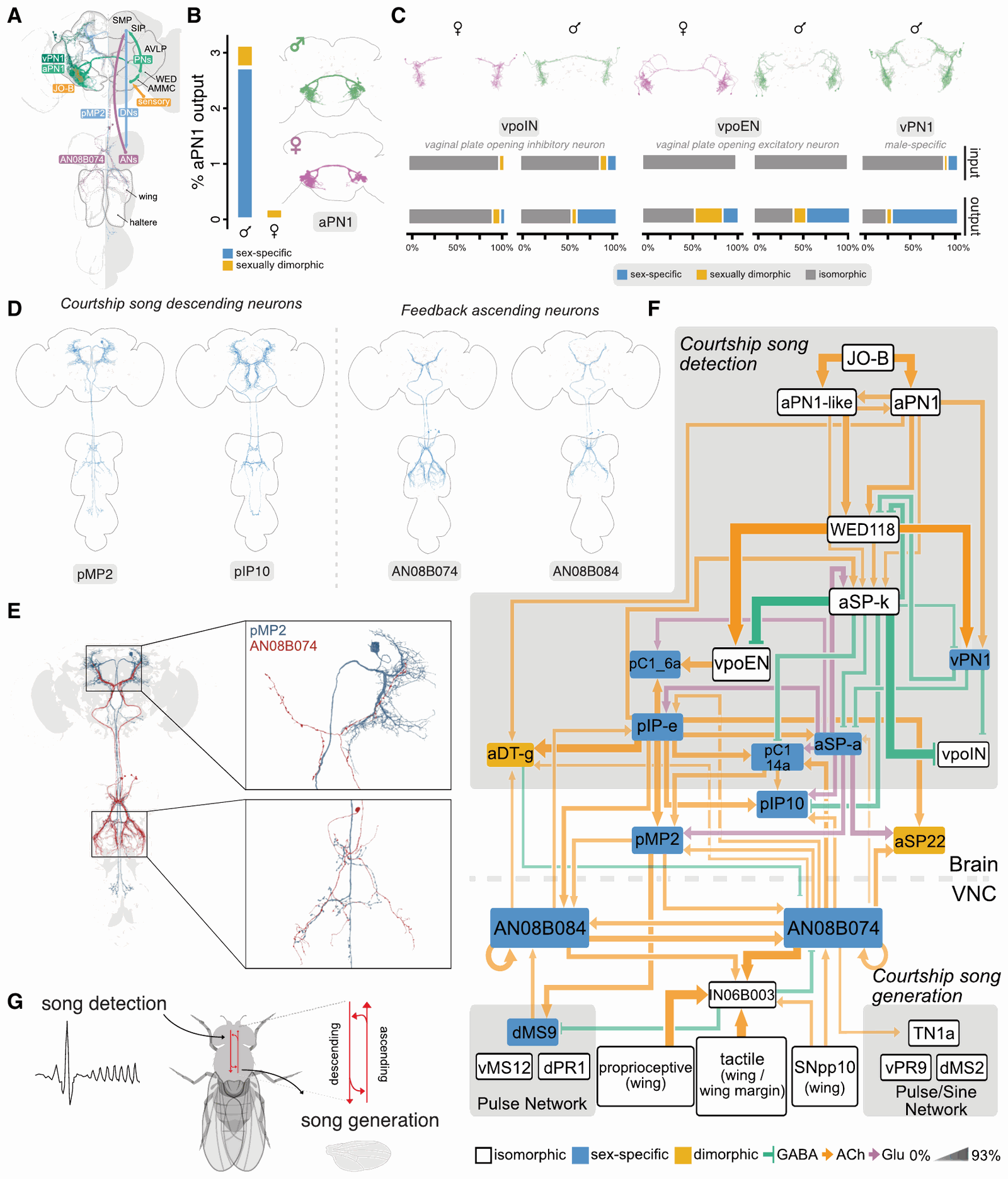
The clearest example needs both halves. Male flies sing by vibrating a wing. Song generation lives in the nerve cord; song detection starts at the antenna and runs through the brain. Extending earlier work, the paper traces two male-specific ascending neuron types, AN08B074 and AN08B084, that carry information from the nerve cord's song circuits up to fru+ song-detection neurons in the brain, and directly to pIP10 and pMP2, the descending neurons that drive song. The connections run both ways, closing a loop from singing back to hearing. The authors suggest it may act as an efference copy, letting a male tell his own song from another's. The function is their hypothesis; the loop is the data.
Sexual dimorphism, counted
The paper sorts every matched cell type into one of three bins by comparing the male CNS with FlyWire's female brain (and the hemibrain):
- Isomorphic: male and female neurons of the type intermingle in morphology and connectivity.
- Dimorphic: the type exists in both sexes, but its neurons separate by sex, with consistent differences in shape or wiring.
- Sex-specific: found in one sex only.
A candidate counts only if it is consistent left and right, in number and morphology. That rule is what makes one specimen per sex usable.
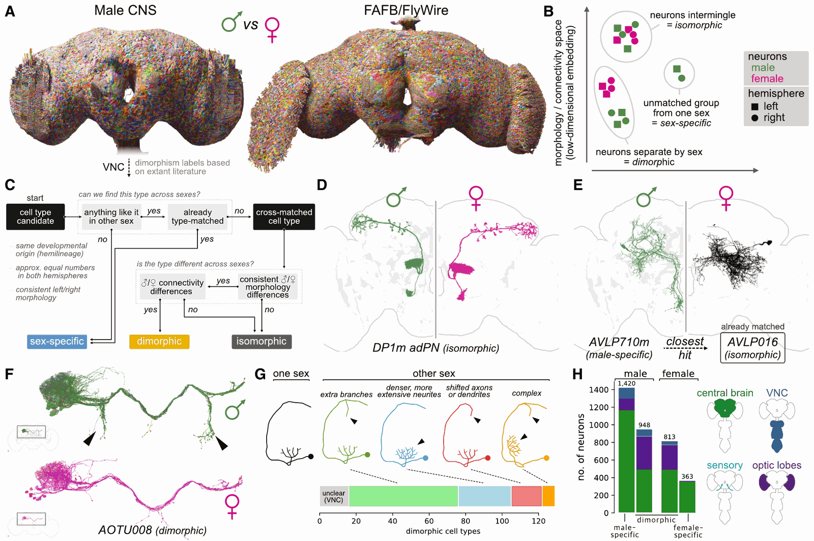
At the type level: 8,069 isomorphic, 138 dimorphic, 289 male-specific and 71 female-specific (reported). At the neuron level: 1,420 male-specific neurons in the male CNS against 363 female-specific neurons in FlyWire, and 948 dimorphic neurons in the male matching 813 in the female (reported). The authors put the dimorphic mismatch down to variability, especially in optic-lobe columns: about 900 in this male, about 800 in FlyWire.
Where are they? Mostly not near the senses:
- In the male central brain, 3.4% of neurons are male-specific and 1.4% dimorphic, 4.8% in total. The female figure is 2.4% (reported).
- In the male visual system, 0.1% are sex-specific and 0.3% dimorphic. Of 335 visual projection neuron types, exactly one, LoVP92, is male-specific (reported).
- A graph traversal assigns every neuron a layer counted from the sensory neurons. Isomorphic neurons average layer 3.9; sex-specific 4.3; dimorphic 4.5. Layers 1–3 hold 18.5% of isomorphic neurons but only 2.5% of male-specific ones (reported).
- A further 6.9% of male central-brain neurons (1.9% in females) look the same in both sexes but take part in dimorphic connections: "secondarily dimorphic" (reported).
The paper's reading: sex differences mainly modify integrative, decision-making areas, while sensory detection and the motor interface stay more constant.
Development. About a quarter of the roughly 200 hemilineages that build the central brain produce dimorphic or sex-specific neurons, but just eight produce more than half of them (reported). One example is the DM4 dorsal lineage: 349 neurons in the male against 202 in the female, or 198 against 201 once sex-specific neurons are excluded (reported). The surplus is the sex-specific cells, not a bigger lineage (reasoned).
Genes. Sex-specific splicing of two transcription factors, fruitless (fru) and doublesex (dsx), shapes how fly neurons develop in each sex. Matching light-microscopy expression patterns to EM neurons gave 4,858 fru neurons (2,804 high confidence) and 412 dsx neurons (331 high confidence), 258 expressing both (reported). In the central brain, 90.4% of male-specific neurons are fru+ or dsx+, against 60.3% of dimorphic and 7.4% of isomorphic ones (reported). So 39.7% of dimorphic neurons carry neither label; the authors blame incomplete expression data in part and leave room for other mechanisms.
A circuit. LoVP92, the one male-specific visual projection type, samples the frontal visual field. Male-specific and dimorphic partners make up 18% of its inputs and 43% of its outputs (reported). One route runs through the male-specific inhibitory interneuron VES200m onto DNg13, a coarse-steering descending neuron. The authors propose that with a female straight ahead, LoVP92 suppresses coarse steering so finer midline tracking takes over, and compare it to the "love spot" larger flies use to track mates. A hypothesis from wiring, labelled as one.
How much to trust n = 1. One male against one or two female brains is a small sample. The hemibrain female at least matches this male's age and genotype: five days old, from a Canton S G1 × w1118 cross (reported in each paper). The paper sizes the risk: developmental errors drop under 1% of neurons per hemisphere, so if the sides fail independently, the chance of missing a whole cell type is under 0.01%, about 3 pairs of central-brain neurons, against more than 1,000 male-specific neurons (reported). Nerve-cord dimorphism labels still come from the literature, because no female nerve cord is fully proofread; the paper expects the female BANC dataset to fill that gap once its proofreading and joint typing mature.
Using it
The data is CC-BY and the paper CC BY 4.0. Everything hangs off the MaleCNS landing page:
- Browse: neuPrint (dataset
male-cns:v1.0) for connectivity; a Neuroglancer scene with EM, segmentation, synapses and FlyWire and hemibrain meshes in male-CNS space; Clio; a Cell Type Explorer and a Dimorphism Explorer. NeuronBridge matches neurons to light-microscopy images of driver lines. - Query from code:
neuprint-pythonor R'sneuprintr, with a neuPrint account token.navisadds skeletons and meshes;coconatflycompares across FlyWire, hemibrain, MANC, BANC and the male CNS. - Bulk download: Feather tables under
gs://flyem-male-cns/v1.0/connectome-data/flat-connectome/: neuron annotations 13 MB, per-neuron transmitter predictions 42 MB, segment-to-segment weights 1.1 GB, synaptic partner pairs 6.8 GB, synapse points 12.7 GB (reported). SWC skeletons and the neo4j database behind neuPrint are there too. - Analysis code for the paper: flyconnectome/2025malecns.
The dataset's documented starting point, one descending neuron type and its partners. I did not run it; it needs a token.
from neuprint import Client, fetch_neurons, fetch_adjacencies

client = Client("https://neuprint.janelia.org", dataset="male-cns:v1.0", token=token)
neurons, syndist = fetch_neurons("DNge104")                   # annotations, per-neuropil synapses
outgoing, _ = fetch_adjacencies("DNge104")                    # its downstream partners
incoming, _ = fetch_adjacencies(None, "DNge104")              # its upstream partners
Two practical notes. Decide early which synapse count you mean (T-bars, postsynapses, proofread connections, thresholded edges); your table will not match a headline. And use consensusNt for transmitter identity, which the paper recommends: it folds in experimental ground truth and marks weakly validated transmitters unclear.
What it does not tell you
- Function. Synapse counts are not synaptic strengths; functional experiments have to confirm what the wiring predicts.
- Every connection. 40.1% of detected connections have both ends proofread.
- Transmitters with certainty. A classifier over 7 classes predicts them; two are marked unclear in the consensus.
- Variation. One male, one age, one moment. The authors note experience modifies even innate dimorphic behaviours; a snapshot cannot show where.
- The female nerve cord. The sex comparison is brain-only until one is finished.
What it does give you is the first male fly nervous system you can follow from eye to leg, neuron by neuron, with the pipeline's error bars published beside it. The expensive stage is still people: 44 person-years, 41% of it linking small fragments. Google's post makes the same point: cutting manual correction is what makes bigger brains affordable. AutoProof, trained on proofreaders' own decisions, already did about 4 person-years of that work here.